Difference between wedge smear and thick drop for blood analysis
The difference between a wedge smear and a thick drop is the amount of blood and the method of spreading the blood on the slide. The two techniques have different purposes.
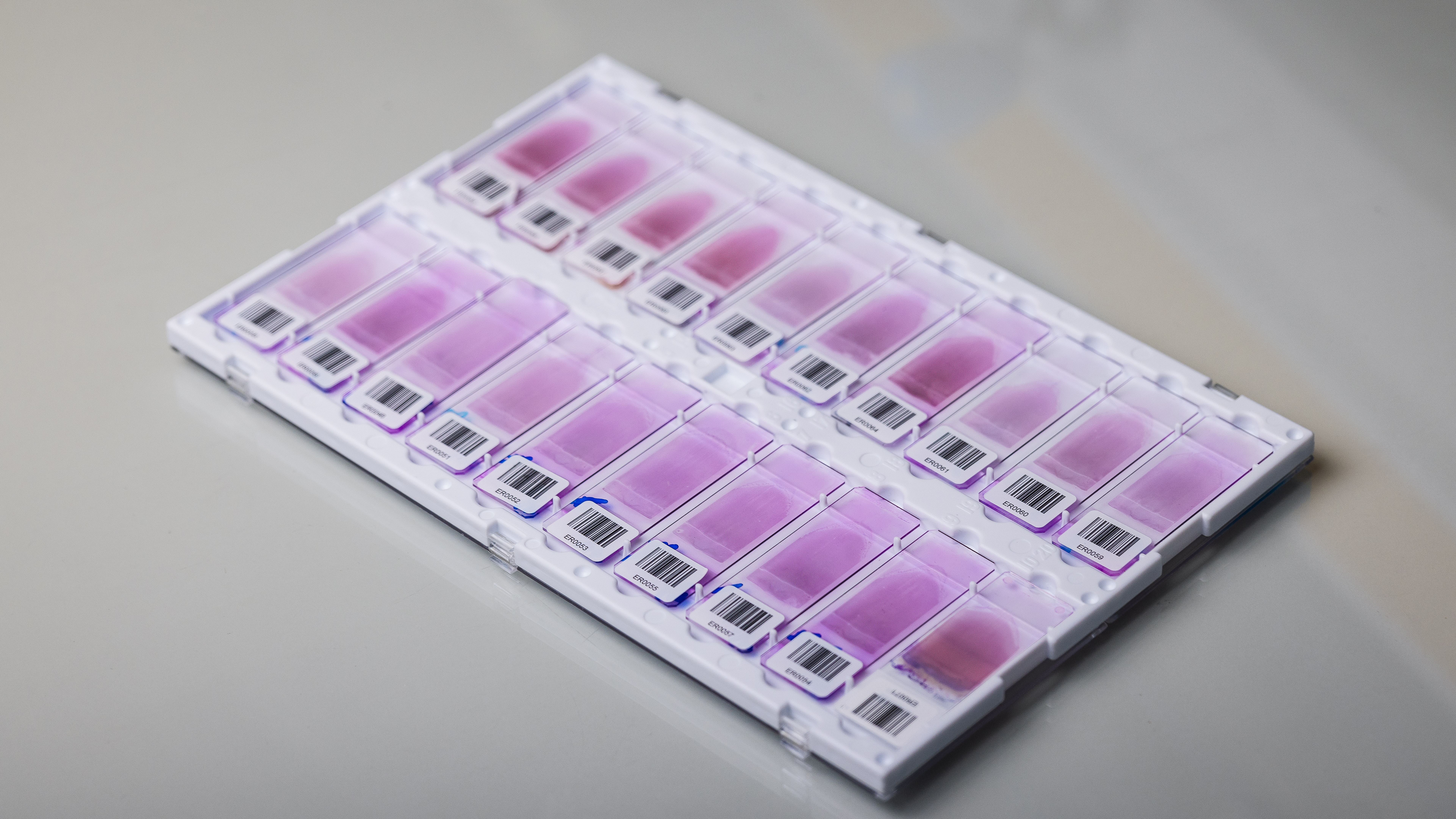
Wedge Smear
What is a wedge smear?
A wedge smear is made by spreading a small amount of blood on a microscope slide. The goal is to obtain a good reading area, or monolayer, where the blood cells are evenly distributed. The size and shape of the monolayer area is important for assessing the morphology of the blood cells.
How to make a wedge smear:
- Mix the EDTA blood tube and place 5-7 µL of blood near the frosted end of the slide.
- Use a second slide as a spreader blade and place it with a 25-45° angle in front of the blood drop and smoothly draw back.
- The blood spread along the spreader blade by capillary action. Then push the spreader blade in a smooth steady motion so that a thin film of blood is spread over the slide. A good-quality wedge smear shows a gradual decrease in thickness.
- It is important that the wedge smear dries quickly and completely before fixing or staining it.
Fixation and Staining
Fixation of the smear is done in either methanol, ethanol, or in the undiluted staining solution used (May-Grünwald, Wright, Wright-Giemsa, or MCDh 1). The fixation step is important to maintain the structures of the cells.
The smear is then stained with a Romanowsky stain: May-Grünwald Giemsa, Wright, Wright-Giemsa, MCDh or 555.
When is a wedge smear used?
A wedge smear is used for:
- WBC differential count
- RBC and WBC Characterization of color, size, shape, and inclusions
- Identification of parasites including its development phase (trophozoites, schizonts, gametocytes) and calculation of the infected cell ratio.
Thick film
What is a thick film?
The thick film is a drop of blood spread on a small area of a microscope slide. The goal is to obtain a concentrated zone of blood and the parasites it contains.
How to make a thick film
- Place several drops of blood in the center of the slide.
- Spread the blood in a circle with a capillary tube or a small stick.
- Allow it to dry, then place the slide in contact with water to allow the lysis of the red blood cells and the release of the hemoglobin.
- The thick film must be completely dry before staining.
Fixation and Staining
The thick film should not be fixed.
Thick films are stained with Giemsa or Leishman stain at pH 7.2.
When is a thick film used?
The thick film is mainly used for diagnosis in parasitology. The concentration of parasites in a thick film is much higher than on a wedge smear so searching for them requires less time, especially in cases where the concentration of parasites is low.